
Synonyms of Pfeiffer Syndrome
- acrocephalosyndactyly, type V
- ACSV
- craniofacial-skeletal-dermatologic syndrome
- Noack syndrome
Subdivisions of Pfeiffer Syndrome
- Pfeiffer syndrome type I
- Pfeiffer syndrome type II
- Pfeiffer syndrome type III
what is Pfeiffer Syndrome?
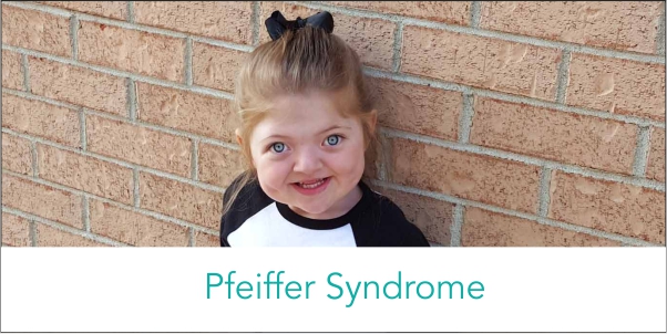
Pfeiffer syndrome is a rare genetic disorder characterized by premature fusion of certain skull bones (craniosynostosis), and abnormally broad and medially deviated thumbs and great toes. Most affected individuals also have differences to their midface (protruding eyes) and conductive hearing loss. Three forms of Pfeiffer syndrome are recognized, of which types II and III are the more serious. Pfeiffer syndrome is an autosomal dominant condition associated with mutations in the genes fibroblast growth factor receptor-2 (FGFR2) and fibroblast growth factor receptor-1 (FGFR1). Pfeiffer syndrome is now known to be a member of a group of conditions caused by mutations in the FGFR genes including Apert syndrome, Crouzon syndrome, Beare-Stevenson syndrome, FGFR2-related isolated coronal synostosis, Jackson-Weiss syndrome, Crouzon syndrome with acanthosis nigricans and Muenke syndrome. (For more information on these conditions, please see the Related Disorders section below.)
Signs & Symptoms of Pfeiffer Syndrome
Infants with Pfeiffer syndrome type I have craniosynostosis that causes the head to appear short and tall (turribrachycephaly). Additional features may include a high, full forehead; underdeveloped midfacial regions (midface hypoplasia); widely spaced eyes (ocular hypertelorism); an underdeveloped upper jaw (hypoplastic maxilla), with a prominent lower jaw; and dental abnormalities. Intelligence is usually normal.
Pfeiffer syndrome type II is characterized by a more severe form of craniosynostosis (Cloverleaf skull), with more severe hand and foot anomalies and additional malformations of the limbs. In infants with Pfeiffer syndrome type II, premature closure of the fibrous joints (cranial sutures) between several bones in the skull causes the skull to have a “tri-lobed” appearance (cloverleaf skull deformity, or Kleeblattschadel type craniosynostosis). In addition, this form of craniosynostosis is often associated with hydrocephalus, a condition in which the normal flow of cerebrospinal fluid (CSF) is altered, leading to abnormal widening (dilatation) of the spaces within the brain (ventricles) causing accumulation of CSF in the skull and increased pressure on the brain. Characteristic craniofacial features associated with Pfeiffer syndrome type II may include an abnormally high, broad forehead; severe protrusion of the eyes (ocular proptosis); an unusually flat middle portion of the face (midface hypoplasia); a “beak-shaped” nose; and downwardly displaced ears. Affected infants may also exhibit abnormal fixation and lack of mobility (ankylosis) of the elbow joints and/or, in some cases, various malformations of certain internal organs in the abdomen (visceral anomalies). In addition, infants with Pfeiffer syndrome type II often experience impaired mental development and neurological problems due to severe involvement of the brain, and/or hypoxia due to problems with breathing. Without appropriate treatment, the physical abnormalities associated with the disorder may lead to life-threatening complications during infancy.
Individuals with Pfeiffer syndrome type III have symptoms and findings similar to those present in Pfeiffer syndrome type II, with the exception of the cloverleaf skull deformity. Additional characteristics associated with Pfeiffer syndrome type III include a shortened base of the skull (anterior cranial base); the abnormal presence of certain teeth at birth (natal teeth); severe protrusion of the eyes (ocular proptosis) due to abnormal shallowness of the bony cavities that accommodate the eyeballs (orbit); and/or various malformations of certain internal organs in the abdominal area (visceral anomalies). As in type II, individuals with Pfeiffer syndrome type III often experience impaired mental development and severe neurological problems and may develop potentially life-threatening complications early in life without appropriate treatment.
Causes of Pfeiffer Syndrome
Pfeiffer syndrome is an autosomal dominant genetic disorder. Dominant genetic disorders occur when only a single copy of an abnormal gene is necessary to cause a particular disease. The abnormal gene can be inherited from either parent or can be the result of a new mutation (gene change) in the affected individual. Essentially all cases of Pfeiffer syndrome type II and type III have resulted from new mutations. Advanced paternal age is associated with an increased risk for new mutations for Pfeiffer syndrome. The risk of passing the abnormal gene from an affected parent to offspring is 50% for each pregnancy. The risk is the same for males and females.
Pfeiffer syndrome type I is associated with mutations in FGFR1 and FGFR2. Pfeiffer syndrome type II and type III are associated with mutations in FGFR2.
Affected Populations
The incidence of all types of Pfeiffer syndrome is approximately 1/100,000.
Related Disorders
Apert syndrome is a rare genetic disorder that is apparent at birth (congenital). The disorder is characterized by distinctive malformations of the head that lead to distinctive facial features. In addition, the hands and/or feet may be webbed (syndactyly) and in some cases, mental retardation may also be present. Babies born with Apert syndrome have fibrous joints between bones of the skull (sutures) that close prematurely (craniosynostosis). The pressure of continued brain growth distorts various bones of the skull and the face. The skull is forced into one of several characteristic shapes. Often the head appears abnormally pointed at the top (acrocephaly). The distortion of the skull plates create changes in the facial bones leading to characteristic facial abnormalities, such as widely spaced eyes (ocular hypertelorism), abnormal protrusion of the eyes (exophthalmos), underdevelopment of midfacial regions (midface hypoplasia), and/or a narrow roof of the mouth (palate).
Malformations of the hands and feet may include unusually broad thumbs and great toes, short fingers, and/or partial to complete fusion (syndactyly) of certain fingers and toes (digits). Most commonly, there is complete fusion of bones within the second to the fourth fingers and the presence of a single common nail (“mitten-like” syndactyly). Apert syndrome is an autosomal dominant genetic condition associated with mutations in FGFR2. (For more information on this disorder, choose “Apert” as your search term in the Rare Disease Database.)
Thanks for reading this content of Dr. Sheibani Nia’s website, The best orthodontist in Tehran.

بدون نظر